pic

Ultrasound

Ultrasound services located in Austin Texas


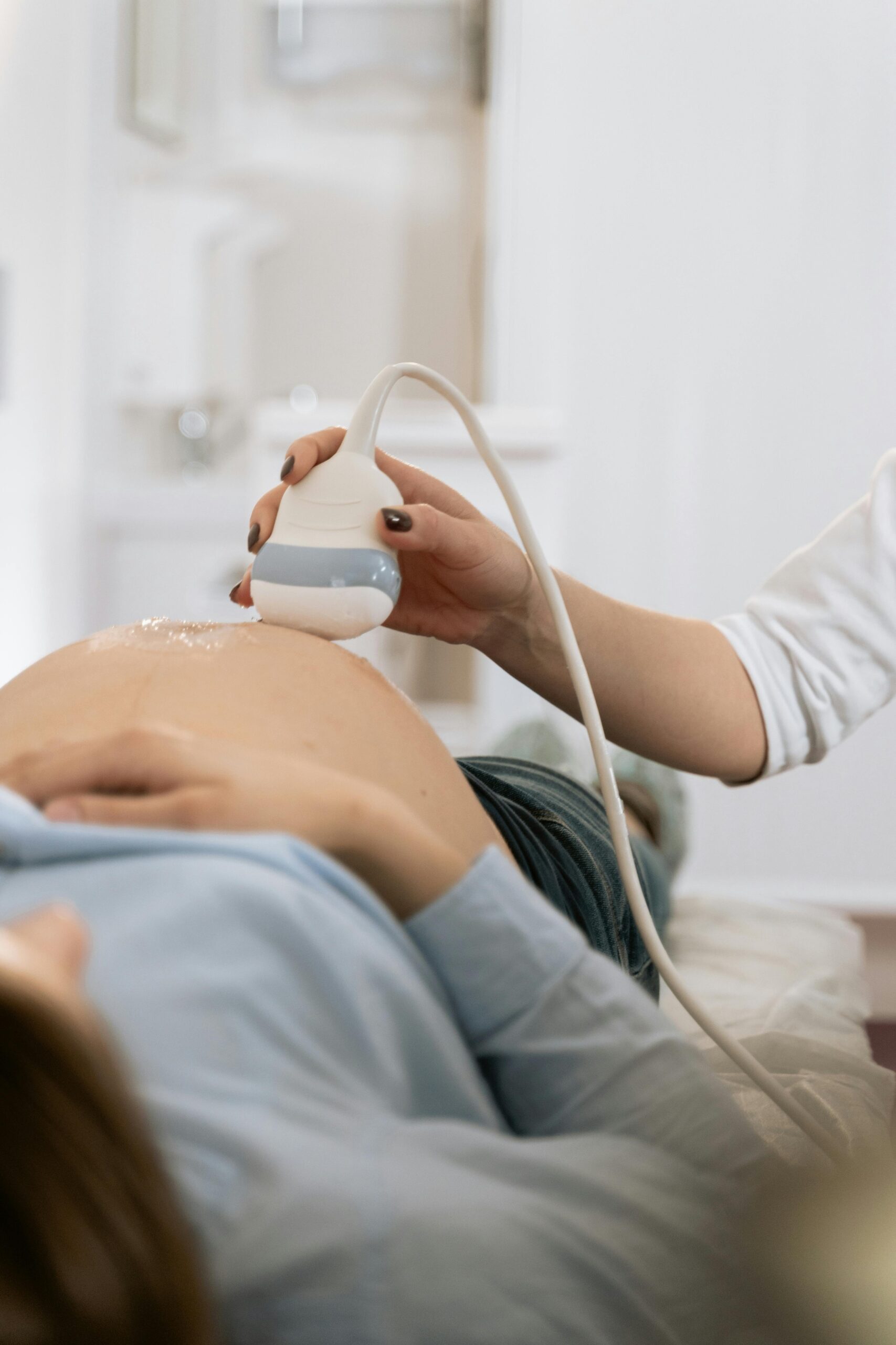
Ultrasound evaluations play an important role in pregnancy, and they have many other applications as well. Located in the heart of Austin, Texas, Austin Area Ob-Gyn & Fertility uses the most advanced ultrasound technology and techniques to provide patients with the most accurate results and optimal care.

Ultrasound Q & A

What is an ultrasound?

An ultrasound exam is a diagnostic imaging exam that uses sound waves to create highly detailed images of organs and other soft tissues, as well as evaluate ongoing processes as they occur. Ultrasounds can produce both still images and videos that can be viewed in real time as well as being stored for evaluation in the future. Unlike x-rays and CT scans, ultrasounds do not use radiation so they're safe for use during pregnancy.

When are ultrasounds used in gynecology?

In addition to their use in monitoring fetal development during pregnancy, ultrasounds also can be used to:

  • assess the pelvic structure and the health of the placenta during pregnancy
  • diagnose causes of some symptoms including vaginal pain and abnormal vaginal bleeding
  • look for tumors, cysts and fibroids
  • assess the structure and function of the uterus, ovaries and fallopian tubes
  • diagnose some types of incontinence
  • understand the causes of pelvic pain
  • monitor management of some conditions

How are ultrasounds performed?

In gynecology, ultrasounds may be performed in two ways: through the skin of the belly region or transvaginally, using a long wand-shaped device designed to be inserted into the vagina for better imaging of the internal structures. Prior to the test, the patient will need to remove her clothing from the waist down and don a gown or use a drape. During the exam, the patient lies on her back in a darkened room. In an abdominal ultrasound, a warm gel will be applied to the skin over the belly and a handheld device called a transducer will be pressed against the skin and passed over it. As the transducer moves across the skin, sound waves will be transmitted through the skin to the interior of the body where they will bounce off organs and other structures and send data back to the ultrasound machine. The machine will interpret the data and produce images on a special viewing monitor. In a transvaginal ultrasound, the transducer wand is lubricated and gently inserted into the vagina rather than being placed on top of the belly.

*Individual Results May Vary